Photo Talk - Winners
Joint Winners
1. TRICK or TREAT – Dr. Rasna Bhanuman
Congenital Ectropion Uveae and Secondary Glaucoma Dr. Shwetha Iyer
2. The Spherical Secret: A Glimpse into Spherophakia Dr. Rashmi Krishnamurthy
Bubble Trouble Dr. Sharmila R
Dr. Archana Singaram
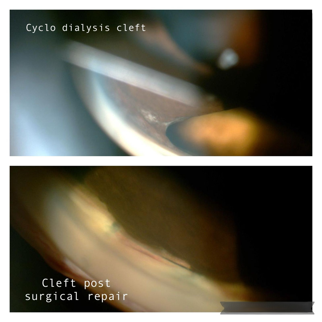
Fixing a Cyclodialysis Cleft
Single mirror gonioscopic image (top image) of a cyclo dialysis cleft with exposed suprachoroidal space that occurred post pars plana vitrectomy port insertion and closure of cleft (bottom image) post ab externo cleft repair.
Dr. Avneet Kaur
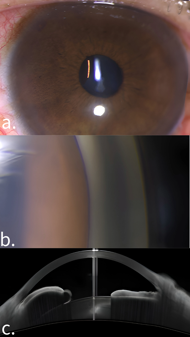
Through the Pupil: Revealing the Mystery of an Iris Cyst
A. Slit lamp photograph showing iris cyst peeking through the pupil
B. Gonioscopic view of the angle narrowing caused by Iris Cyst
C. Anterior Segment OCT showing iris cyst
Dr. Balam Pradeep
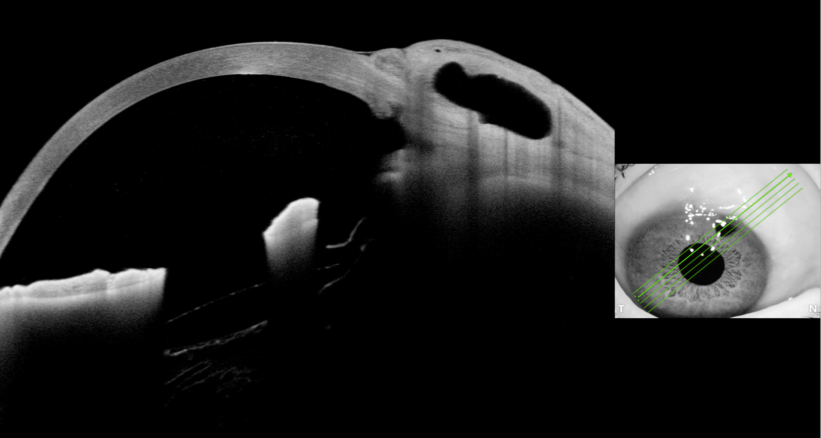
Imaging of Scleral Flap Patency and Bleb Analysis Using Anterior Segment Optical Coherence Tomography
Imaging Showing The Surgical Patent Iridectomy,Patent Sclerostomy With Scleral Track Under The Partial Thickness Scleral Flap and A Well Formed Bleb Morphology With Good Filtration of Aqueous Is Noted.
Dr. Gazella Bruce
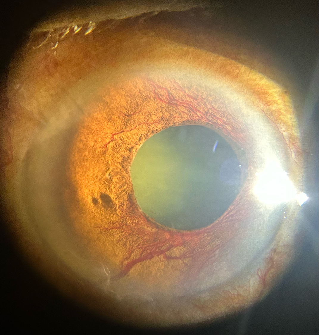
Neovascular Glaucoma with Neovascularization of Iris in Diabetic Retinopathy
The tree of life may have branches growing into the light, But don't let the tree in glaucoma and diabetes steal your sight
Dr. Jignesh Jethva
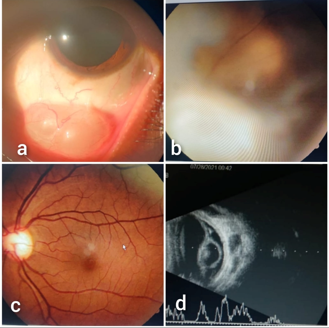
Patient was Known Case of Neurocysticercosis with Occular Involvement..
Figure a) left eye subconjunctival cyst
Figure b) cyst in vitreous cavity in right eye
Figure c) 0.8 CDR ( Open angle glaucoma) with barring and bayoneting sign positive
Figure d) USG of right eye suggest of Cyst in vitreous
Dr. John Davis
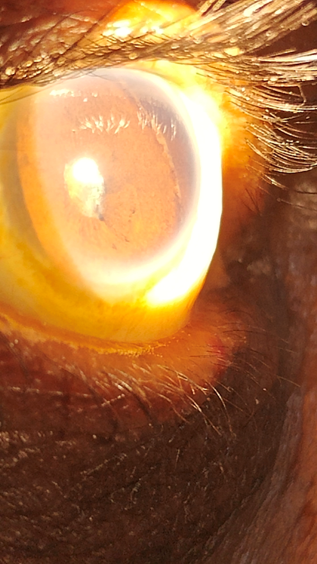
Hidden Angles Come to Light,Cyclodialysis in Clear Sight
In a patient with mature traumatic cataract, nasal iridocorneal angle is seen directly when viewed from the temporal side of the eye due to a large cyclodialysis cleft of 200 degrees. Cataract surgery with PCIOL was later done successfully in this patient
Dr. Manju M
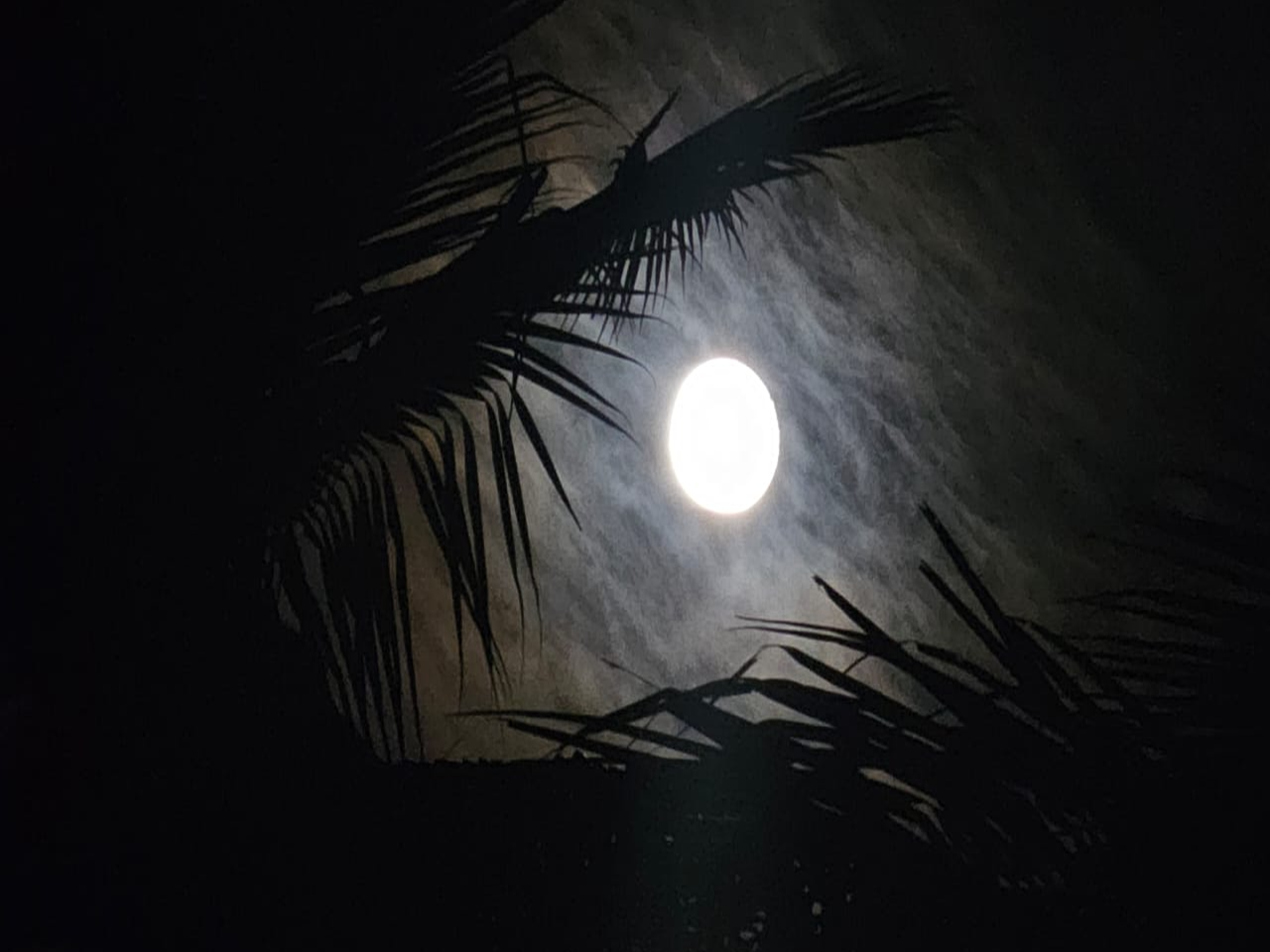
When Shadows Steal Sight
This image beautifully captures the essence of glaucoma—a silent thief of sight that creeps in like the shadows surrounding the moon. The bright moon represents clarity and vision, while the encroaching darkness mirrors the gradual loss of sight caused by the disease. Just as light struggles to break through the clouds, individuals with glaucoma fight against the progressive narrowing of their visual field. However, with early detection and proper treatment, vision can be preserved, allowing the light to emerge from the darkness, much like the tale of vision restored.
Dr. Parul
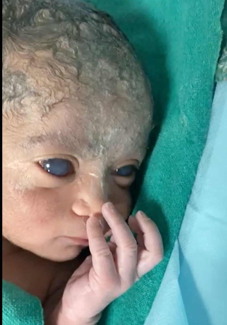
Hope in Every Blink: A Newborn's Fight Against Glaucoma
Dr. Rashmi Krishnamurthy
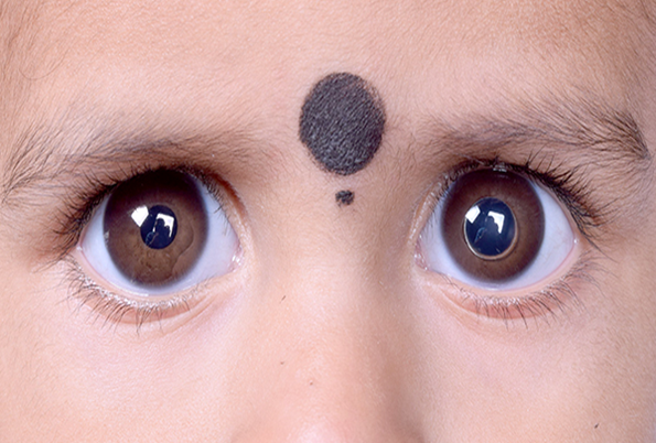
The Spherical Secret: A Glimpse into Spherophakia
A striking clinical image of a 2-year-old girl with bilateral Spherophakia and spontaneous anterior lens dislocation in the left eye. As the lens is the primary pathology, lens explantation is the main treatment, with glaucoma surgery considered only for uncontrolled IOP post-explantation.
Dr. Rasna Bhanuman
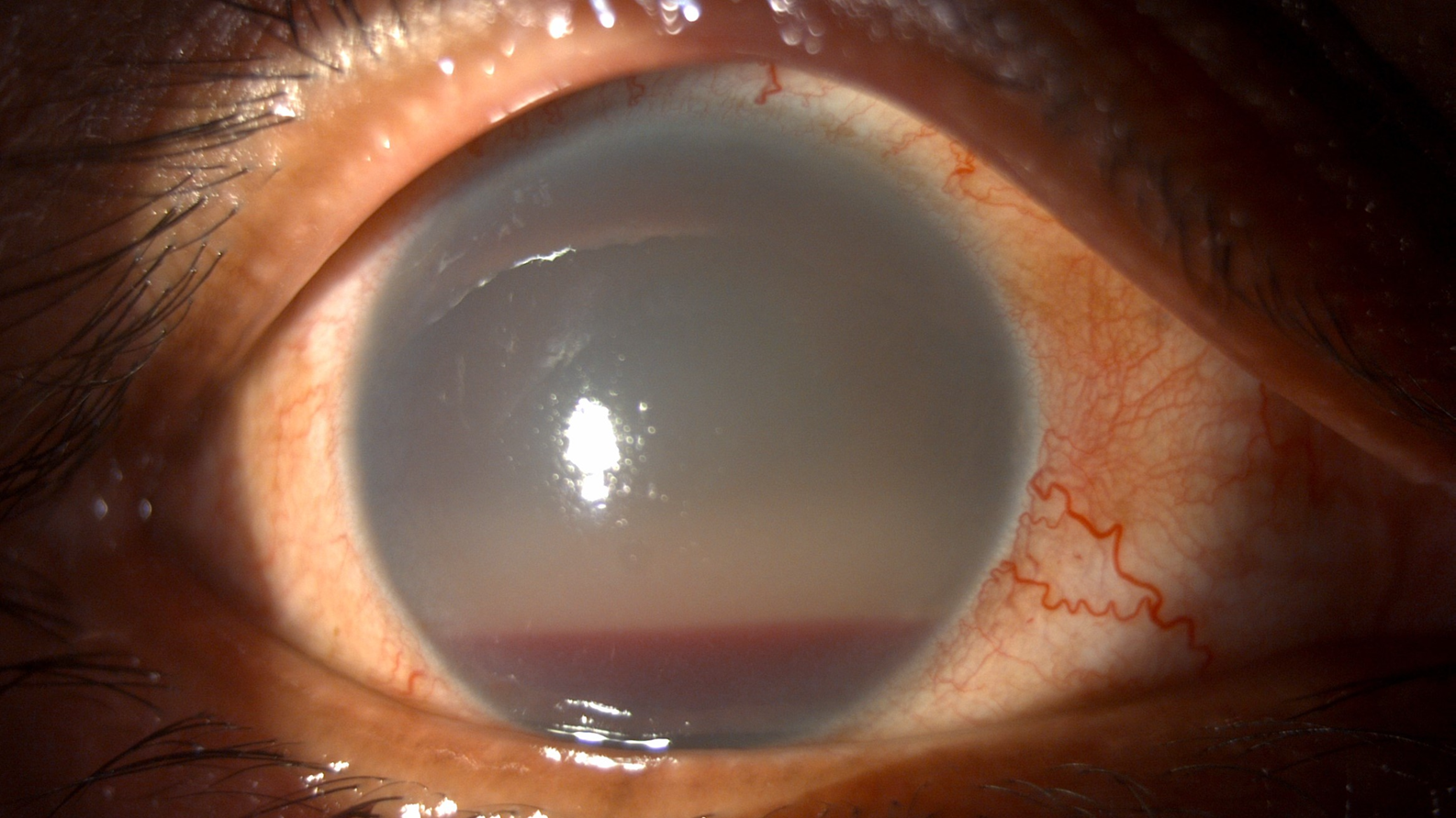
Trick or Treat
A trauma tale where blood and fluid meet. Slit lamp photo of an evolving candy stripe hyphema post trauma ,seen in Ghost cell glaucoma.
Dr. Saradha Ravi
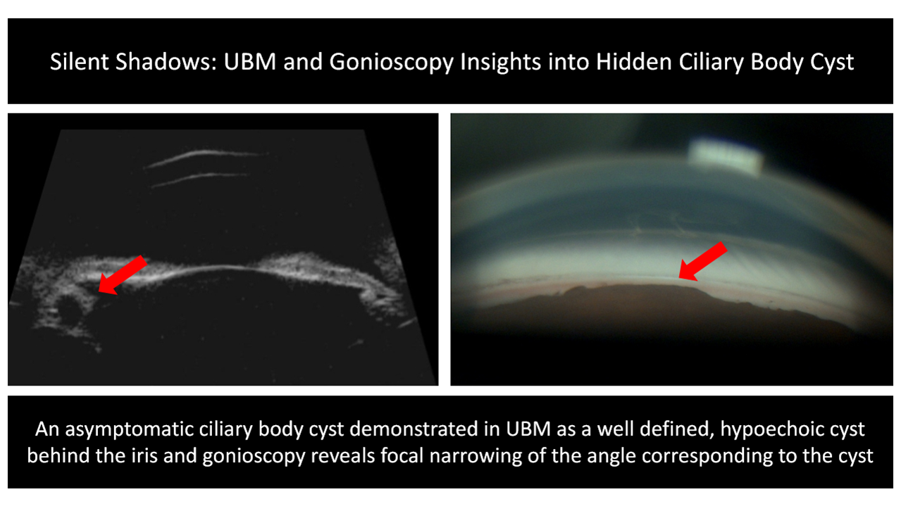
Silent Shadows: UBM and Gonioscopy Insights into Hidden Ciliary Body Cyst
An asymptomatic ciliary body cyst demonstrated in UBM as a well-defined, hypoechoic cyst behind the iris, and gonioscopy revealed focal narrowing of the angles corresponding to the cyst.
Dr. Sharmila R
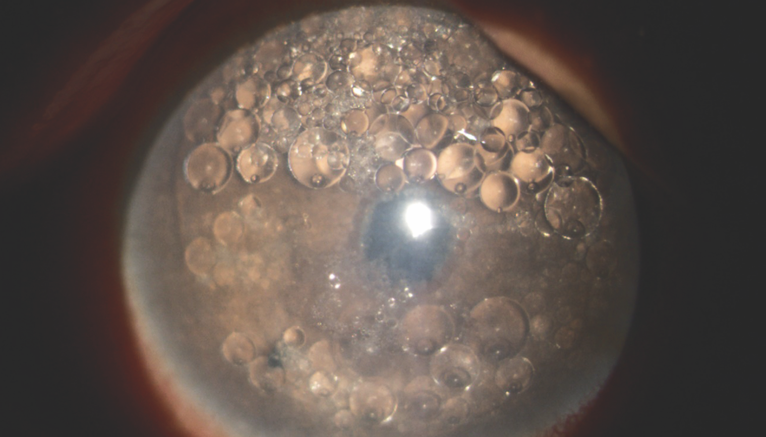
Bubble Trouble
Silicon oil is a vitreous substitute commonly used in Vitreo-retinal surgeries which may cause secondary open or closed angle glaucoma.
Dr. Shwetha Iyer
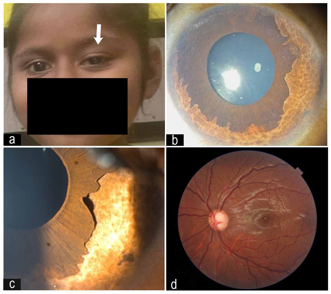
Congenital Ectropion Uveae and Secondary Glaucoma
This is a unique case of an asymptomatic 6 year old child with unilateral advanced glaucoma with intraocular pressure(IOP) of 52mmHg, secondary to Congenital Ectropion Uveae who was referred to us for strabismus evaluation from a school screening program. She had no systemic associations. She underwent Combined Trabeculotomy+Trabeculectomy with Mitomycin-C 0.2mg/ml. Post surgery, her vision improved and IOP lowered to mid teens and remains stable until now. Careful periodic examination and early surgical intervention are required to preserve the productive vision in these children who tend to present late to hospital due to unilateral affliction of the disease.
a. Left eye upper lid ptosis (white arrow)
b. Slit lamp image showing 360-degree ectropion uveae
c. Magnified image of 1b
d. Fundus image showing advanced optic disc cupping.
Dr. Sivani Kodali
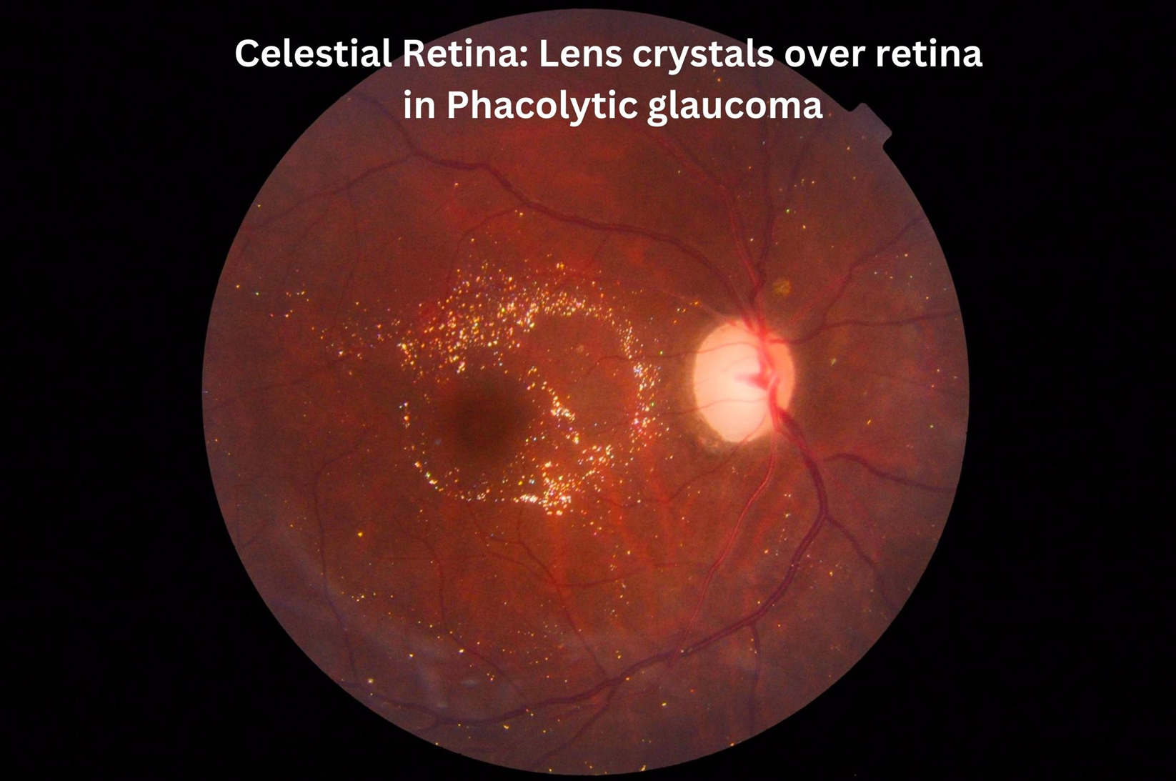
Celestial Retina : Lens Crystals Over Retina in Phacolytic Glaucoma
We observed lens derived crystals on the retina after cataract surgery in an eye with phacolytic glaucoma, resembling a starry pattern- Celestial retina. Likely from degenerating lens material
Dr. Sumitha
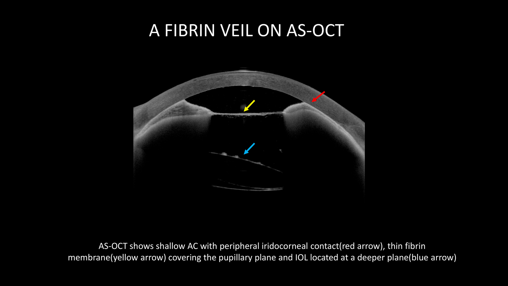
A Fibrin Veil on as - OCT
AS-OCT shows shallow AC with peripheral iridocorneal contact (red arrow), thin fibrin membrane (yellow arrow) covering the pupillary plane and IOL located at a deeper plane(blue arrow)
